Translate this page into:
Hyperpigmentation in a child as a clue to chikungunya
*Corresponding author: Sanjanaa Srinivasa, Department of Dermatology, St John’s Medical College, Bengaluru, Karnataka, India. sanjanaa.srinivasa@stjohns.in
-
Received: ,
Accepted: ,
How to cite this article: Srinivasa S, Ballal S. Hyperpigmentation in a child as a clue to chikungunya. Karnataka Paediatr J. 2024;39:117-8. doi: 10.25259/KPJ_27_2024
Dear Editor,
A 2-month-old male child born of non-consanguineous marriage presented with high-grade, intermittent fever for 1 month along with history of reduced feeding and lethargy for 2 days. The birth weight of the child was 2.5 kg. The mother observed pigmentation initially appearing over the face, which progressively extended to involve the rest of the body for 20 days. All milestones were appropriate for age.
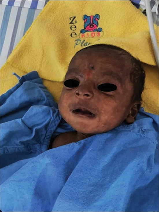
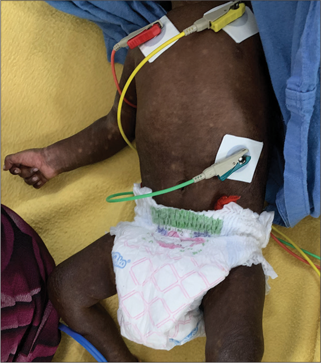
On examination, diffuse macular brownish-black pigmentation was observed over the nose, perioral area, periphery of the face, trunk, and extremities [Figures 1 and 2]. Differentials thought of were Vitamin B12 deficiency, Addison’s disease, congenital adrenal hyperplasia (CAH), and chikungunya.

- Two-month-old male, with chikungunya, with brown black macules and patches over nose, perioral area, forehead, and sides of the face.

- Patchy brownish-black hyperpigmentation over the trunk and extremities.
Routine investigations showed leukocytosis. In view of hyperpigmentation, Vitamin B12 deficiency and CAH were ruled out because Vitamin B12 and serum cortisol were normal. In view of fever, lethargy, and leucocytosis, cerebrospinal fluid (CSF) analysis was performed to rule out meningitis. CSF analysis was normal, with no evidence of meningitis. To rule out chikungunya-induced hyperpigmentation, CSF serology was performed, which tested positive for chikungunya immunoglobulin (IgM). The mother also tested positive for chikungunya IgM; however, she had no antenatal or postnatal history suggestive of chikungunya. This suggests that both the child and mother acquired the infection postnatally through vectors.
The fever and dehydration were attributed to be secondary to sepsis, and the diffuse type of pigmentation was due to chikungunya.
The child was managed conservatively with IV fluids and analgesics, with gradual resolution of hyperpigmentation spontaneously.
Chikungunya is a re-emerging infection caused by Arbovirus, transmitted by Aedes aegypti and Aedes albopictus mosquito. Other rare modes of transmission include blood-borne and vertical transmission in the first or second trimester of pregnancy. A higher risk of vertical transmission occurs when there is a maternal infection within 1 week of delivery, with greater chances of severe disease in the neonate.[1] Pigmentary alterations reported in chikungunya include localised, grey-black, macular hyperpigmentation over the ala of the nose known as chik sign, centrofacial freckle-like/melasma-like macules/patches, flagellate hyperpigmentation, diffuse hyperpigmentation of the face and extremities, mucosal hypermelanosis of the tongue and palate, periorbital hypermelanosis, Addisonian-type palmar pigmentation, and melanonychia.[2]
Generalised pigmentation is more commonly seen in infants compared to centrofacial and neck pigmentation seen in adults. Children more commonly have neurological and dermatological symptoms compared to infected adults.[1]
Pigmentary changes have been attributed to pigment dispersion by the chikungunya virus and post-inflammatory response to the virus.[3]
Other cutaneous manifestations of chikungunya include maculopapular eruption, aphthae like orogenital ulcers, palmar erythema, vesiculobullous lesions, haemorrhagic manifestations, exacerbating of existing dermatosis, telogen effluvium, erythema nodosum, erythema multiforme-like lesions, lichenoid lesions, photosensitivity, lobular panniculitis, lymphoedema, and urticaria.[2]
Chikungunya should be included in the differential diagnosis when evaluating patients presenting with fever and pigmentary changes, particularly in endemic countries. Clinicians should also recognise the diverse cutaneous manifestations associated with chikungunya.
Ethical approval
Institutional review board approval is not required.
Declaration of patient consent
The authors certify that they have obtained all appropriate patient consent.
Conflicts of interest
There are no conflicts of interest.
Use of artificial intelligence (AI)-assisted technology for manuscript preparation
The authors confirm that there was no use of artificial intelligence (AI)-assisted technology for assisting in the writing or editing of the manuscript and no images were manipulated using AI.
Financial support and sponsorship
Nil.
References
- Chikungunya in children: A clinical review. Pediatr Emerg Care. 2018;34:510-5.
- [CrossRef] [PubMed] [Google Scholar]
- Mucocutaneous manifestations of chikungunya fever: An update. Int J Dermatol. 2023;62:1475-84.
- [CrossRef] [PubMed] [Google Scholar]
- Varied cutaneous manifestation of chikungunya fever: A case series. Int J Dermatol. 2017;3:289-92.
- [CrossRef] [Google Scholar]


