
Translate this page into:
A rare presentation of Patau syndrome with complex congenital anomalies - A case report
*Corresponding author: Mini Rajeevan Methalthody, Department of Pediatrics, Kurunji Venkatramana Gowda Medical College, Dakshina Kannada, Karnataka, India. minirajeevanmethalthody@gmail.com
-
Received: ,
Accepted: ,
How to cite this article: Methalthody MR, Rudrappa S. A rare presentation of Patau syndrome with complex congenital anomalies - A case report. Karnataka Paediatr J. doi: 10.25259/KPJ_59_2024
Abstract
Patau syndrome is a rare and complex genetic condition caused by an extra copy of chromosome 13. We report a case of a 3-day-old female infant with Patau syndrome, presenting with multiple congenital anomalies. The genetic study was positive and the infant was managed with supportive care, including oxygen therapy, orogastric feeds, and intravenous antibiotics and discharged with medications for pulmonary artery hypertension and congestive heart failure. This case highlights the importance of early diagnosis and supportive care for infants with Patau syndrome.
Keywords
Genetic anomalies
Patau syndrome
Clefts

INTRODUCTION
Patau syndrome is a rare and complex genetic disorder characterised by the cardinal triad of orofacial clefts, microphthalmia and postaxial polydactyly of the limbs.[1] The incidence of the condition is 1 in 12000–20000 live births worldwide, with a median survival rate of 2.5 days and only 5% of infants surviving beyond 6 months.[2] A systematic review of 60 studies conducted between the period of 2000 and 2021 revealed that the prevalence of Patau syndrome among babies born with congenital anomalies ranged from 0.26 to 5.8%, with a female predominance at birth.[3] More than 90% of the infants die in the 1st month and approximately 1% live beyond 1 year.[4] Trisomy 13 is a rare genetic condition that occurs due to the extra copy of the 13th chromosome; it is associated with a high rate of spontaneous abortion and poor infant survival rate. Infants diagnosed with this exhibit a range of symptoms affecting various parts of the body, with severity levels differing from one individual to another. The common features are mental retardation, congenital heart defects, cleft lip and palate, eye defects, polydactyly, scalp defect, hypotonia and holoprosencephaly.[5]
Patau syndrome occurs when there is an error during mitosis, resulting in an extra copy of chromosome 13; this can occur in three ways: Trisomy 13, Mosaicism and Translocation trisomy 13.[6] The prognosis of this condition is generally poor; therefore, management of Patau syndrome focuses on providing supportive care and alleviating symptoms rather than curing the condition. We are hereby reporting a case of Patau syndrome in a 3-day-old female baby.
CASE REPORT
A 3-day-old female baby [Figure 1] with a birth weight of 2.57 kg was delivered by a caesarean section and was admitted to the intensive care unit, department of paediatrics, due to characteristic features suggestive of Patau syndrome. The baby was born to a 32-year-old non-consanguineous P2L2 mother. The baby cried after suction and stimulation, with an appearance, pulse, grimace, activity, respiration score of 7/10 at 1 min and 8/10 at 5 min. The occipitofrontal circumference was 38 cm, while the body length was 47.4 cm. The baby presented with hurried breathing and bluish discoloration of limbs.
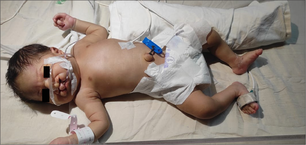
- Patient demonstrating congenital anomalies associated with Patau syndrome.
Oxygen therapy was initiated immediately through nasal prongs, and the baby was started on orogastric feeds and intravenous antibiotics. The comprehensive examination revealed multiple congenital anomalies including aplasia cutis [Figure 2a], trigonocephaly, hypotelorism, upward slant of eye, bulbous nose [Figure 2b], cleft lip and palate [Figure 2c], right post-axial polydactyly, overriding of fingers bilaterally [Figure 2d], anovestibular fistula [Figure 2e], rocker bottom feet, bilateral hallux valgus and hammer toe [Figure 2f]. The diagnostic evaluation revealed a Grade 2 systolic murmur, indicating mild cardiac dysfunction and the septic screen showed positive, indicating a possible infection. Ultrasound of the abdomen revealed normal and neurosonogram showed dilated occipital horns of bilateral lateral ventricles with non-visualisation of the posterior part of the corpus callosum, suggestive of partial corpus callosum agenesis. 2D Echocardiogram showed dilated right atrium/right ventricle, severe pulmonary artery hypertension, reduced right ventricular function, 5 mm ostium septum Atrial septal defect with left to right shunt and moderate left ventricular dysfunction. A genetic study (karyotyping) was positive for Patau syndrome and the baby was discharged with sildenafil and furosemide. The infant’s mother received genetic counselling and she was referred for palliative care to assist with home care of the baby and recommended for breastfeeding.
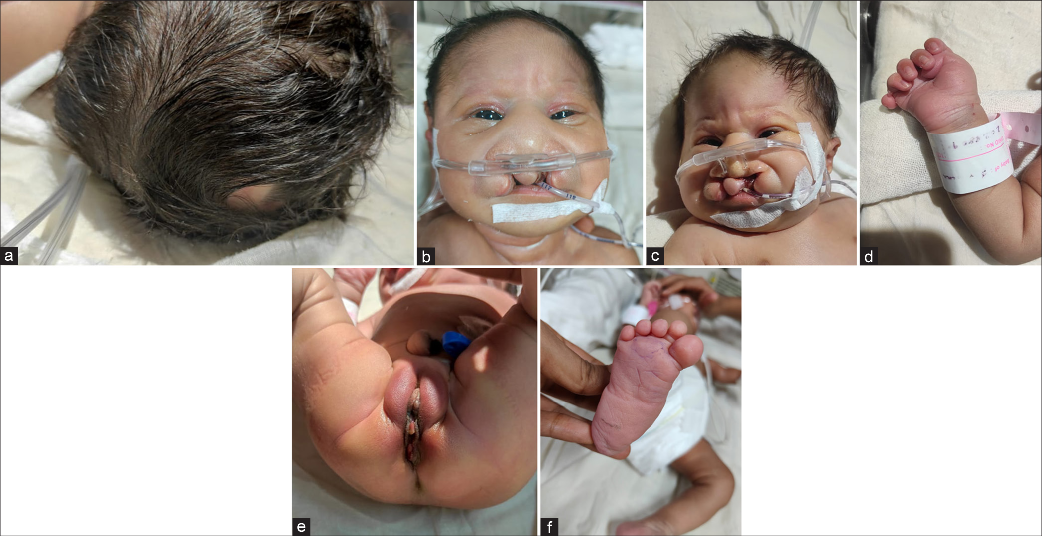
- (a) Aplasia cutis, (b) hypotelorism, upward slant of eye and bulbous nose, (c) cleft lip and palate, (d) right post-axial polydactyly and overriding of fingers bilaterally, (e) anovestibular fistula and (f) rocker bottom feet, bilateral hallux valgus and hammer toe.
DISCUSSION
The present case describes a 3-day-old female infant with Patau syndrome, a rare genetic condition caused by an extra copy of chromosome 13. The infant had several congenital anomalies, including aplasia cutis, trigonocephaly, hypotelorism, polydactyly and complex cardiac defects. Patau syndrome is a rare genetic condition with an average incidence of approximately 1 in 12000–1 in 20000 live births worldwide. Previous studies have reported several cases of Patau syndrome and associated complications. A study published in the American Journal of Paediatrics, using the metropolitan Atlanta congenital defects program, identified 70 live-borns with trisomy 13 and 114 live-born infants with trisomy 18. Median survival time was 7 days (95% confidence interval [CI]: 3–15) for people with trisomy 13 and 14.5 days (95% CI: 8–28) for people with trisomy 18. These infants with Patau syndrome had congenital heart defects, including ventricular septal defects, atrial septal defects and pulmonary artery hypertension.[7] Our case report is consistent with these findings, as the infant had a 5 mm ostium septum ASD with the left to right shunt and severe pulmonary artery hypertension.
A study done by Misanovi et al., reported a case of Patau syndrome having multiple congenital anomalies, including microcephaly, dolichocephaly, microphthalmia and ultrasound changes of the heart and genitourinary system. The case was confirmed cytogenetically with the help of Fluorescent in situ hybridisation techniques.[8] Zhou et al., presented a case of Patau syndrome with an extra copy of chromosome 13 due to nondisjunction during the first meiotic division. This case was confirmed by the FISH technique. The life expectancy of infants with Patau syndrome is very low with a median survival range which is 5–7 days and only 5% of infants surviving beyond 6 months.[9] This low life expectancy is due to the severity of cardiac and cerebral malformations and other multiple congenital anomalies. Patients with Patau syndrome who make it past infancy usually have seizures, severe psychomotor dysfunction and intellectual incapacity. There is no specific treatment for Patau syndrome and it is managed according to the severity of the condition on a case-by-case basis.[10] In our case, the baby was managed with oxygen therapy, orogastric feeds and intravenous antibiotics and was subsequently discharged with sildenafil and furosemide. The outcome for our patient is uncertain; however, with supportive care and management, we hope to improve the quality of life for our patient and family. This case of Patau syndrome highlights the significant challenges in managing this complex condition. Beyond the medical management of the presenting anomalies, a crucial aspect of care involves navigating complex ethical considerations related to the level of intervention and the overall goals of care. Open communication with the family, shared decision-making and ongoing support is essential to ensure the best possible outcome for both the infant and the family. Future case reports and research should focus not only on the clinical features and genetic aspects of Patau syndrome but also on the ethical dimensions of care and the development of best practices for supporting families facing this difficult diagnosis.
CONCLUSION
Patau syndrome can be clinically diagnosed based on the clinical phenotype of the affected infant. However, there is no specific cure for this genetic condition. Referral to a genetic counselling clinic is advocated. In conclusion, our case highlights the importance of early diagnosis of Patau syndrome. Our study also emphasises the significance of supportive care for the affected infant and family that all healthcare providers can offer.
Ethical approval
Institutional Review Board approval is not required.
Declaration of patient consent
The authors certify that they have obtained all appropriate patient consent.
Conflicts of interest
There are no conflicts of interest.
Use of artificial intelligence (AI)-assisted technology for manuscript preparation
The authors confirm that there was no use of artificial intelligence (AI)-assisted technology for assisting in the writing or editing of the manuscript and no images were manipulated using AI.
Financial support and sponsorship: Nil.
References
- Natural history of trisomy 18. Arch Dis Child. 1996;75(1 Suppl):343-5.
- [CrossRef] [PubMed] [Google Scholar]
- Proportion of chromosomal disorders and their patterns among births with congenital anomalies in Africa: A systematic review and meta-analyses. ScientificWorldJournal. 2022;2022:6477596.
- [CrossRef] [PubMed] [Google Scholar]
- Prevalence and pattern of congenital anomalies as seen in a tertiary hospital in Benin city. Int J Innov Sci Res Rev. 2024;6:5713-7.
- [Google Scholar]
- The pathology of Trisomy 13 syndrome-A study of 12 cases. Hum Genet. 1988;80:349-56.
- [CrossRef] [PubMed] [Google Scholar]
- Population-based analyses of mortality in trisomy 13 and trisomy 18. Pediatrics. 2003;111:777-84.
- [CrossRef] [PubMed] [Google Scholar]
- The paternal origin of an extra chromosome 13 from the patient with Patau syndrome. Yi Chuan. 2005;27:349-50.
- [Google Scholar]
- Longevity and Patau syndrome: What determines survival? BMJ Case Rep. 2012;2012:bcr0620114381.
- [CrossRef] [PubMed] [Google Scholar]


