Translate this page into:
Subaponeurotic fluid collection – An unusual cause of scalp swelling in infancy
*Corresponding author: Padmasani Venkat Ramanan, Department of Paediatrics, Sri Ramachandra Institute of Higher Education and Research, Chennai - 600 093, Tamil Nadu, India. padmasani2001@yahoo.com
-
Received: ,
Accepted: ,
How to cite this article: Prasath TS, Gutta MC, Murali A, Ramanan PV. Subaponeurotic fluid collection – An unusual cause of scalp swelling in infancy. Karnataka Paediatr J 2020;35(2):121-3.
Abstract
Subaponeurotic fluid collection (SFC) is a rare cause of scalp swelling in infants that is not well described in textbooks. It can be diagnosed clinically and managed conservatively. We report two infants, who had scalp swelling, with findings suggestive of SFC that resolved spontaneously. We have reviewed literature on the possible mechanisms of fluid collection. We wish to highlight the self-limiting nature of this condition so that unnecessary investigations, referrals, and interventions are avoided.
Keywords
Subaponeurotic fluid collection
Infant
Scalp swelling
INTRODUCTION
Scalp swellings are quite common in the newborn and the causes include cephalhematoma and caput succedaneum. Similar swellings beyond the newborn period cause anxiety among parents and treating pediatricians. Subaponeurotic fluid collection (SFC) as a cause of scalp swelling is not well described in textbooks.[1] It runs a benign course and resolves spontaneously. Awareness about this condition among pediatricians is essential, to avoid unnecessary investigations, referrals, and interventions. We report two infants, who presented scalp swelling, which was diagnosed clinically as SFC and managed conservatively with complete resolution of swelling.
CASE DESCRIPTION
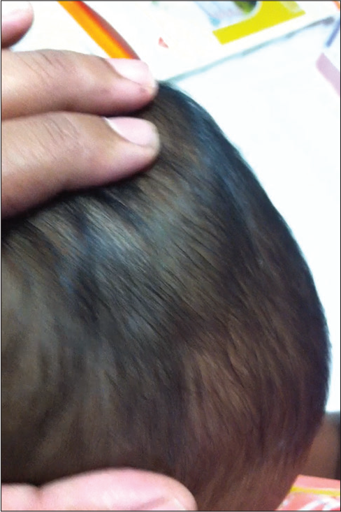
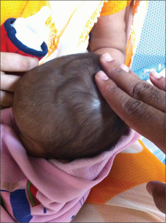
Case 1 was a boy aged 11 weeks, born by normal vaginal delivery, and Case 2, a girl aged 9 weeks born by emergency LSCS – because of arrest of labor. Both presented with an ill-defined scalp swelling of 1-day duration [Figures 1 and 2]. There was no history of forceps or ventouse history of application before delivery or fetal scalp electrode placement. In both infants, there was no bruising, scalp swelling, or cephalhematoma in the immediate newborn period. On examination, both had soft, non-tender, fluctuant swellings not limited by suture lines in the parieto-occipital region. A fluid shift was noted on head movement. The hemoglobin level, platelet count, and clotting profile were normal.

- Scalp swelling in Case 1.

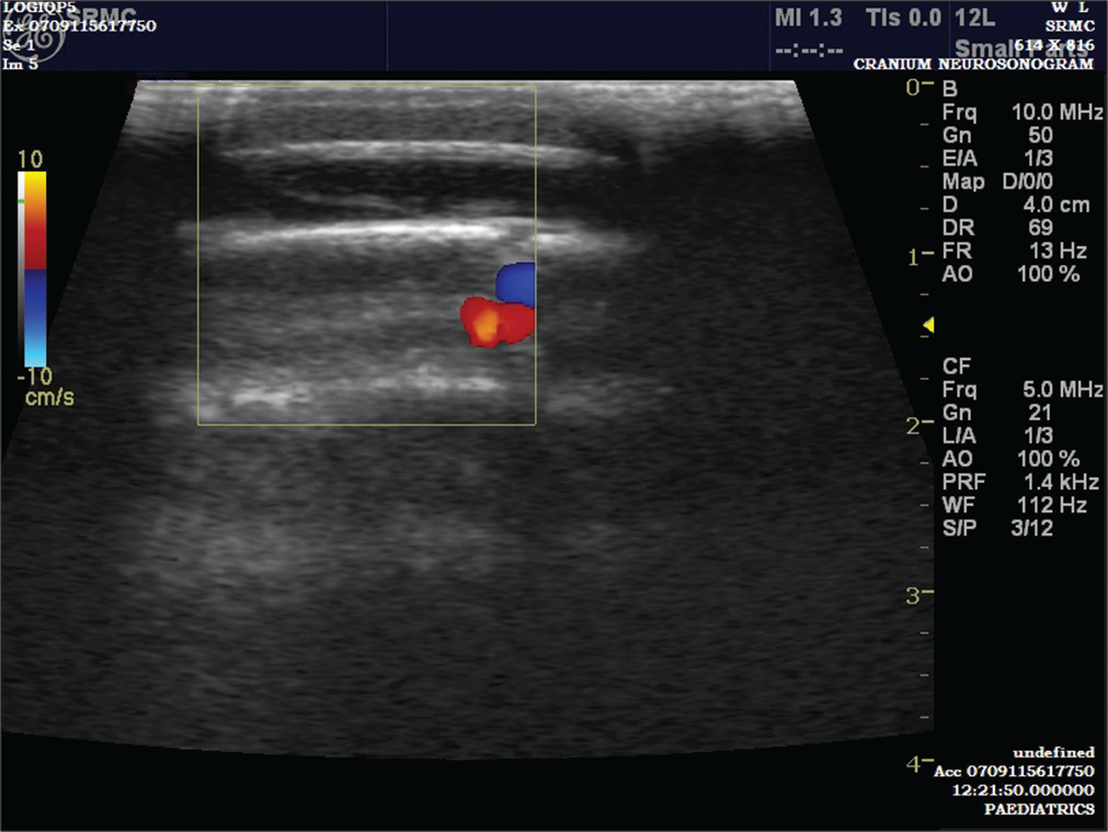
- Ultrasound sonography showing subaponeurotic fluid collection in case 1.
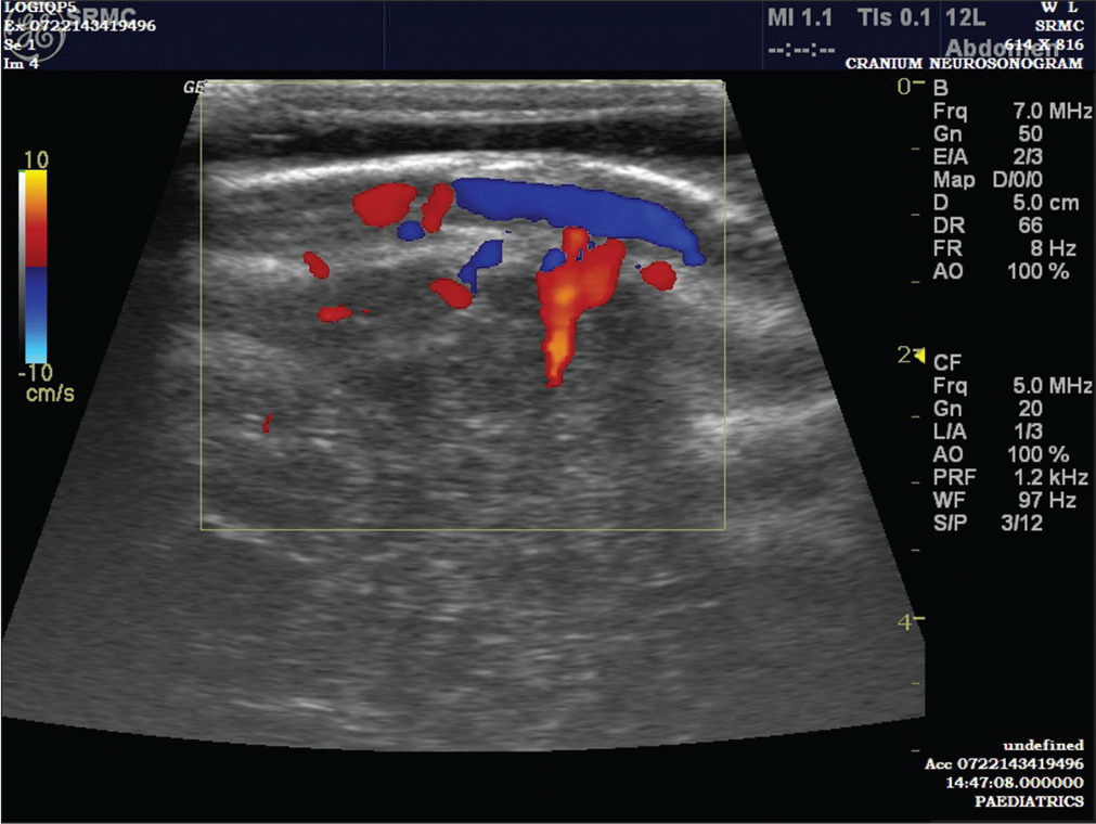
In both, a clinical diagnosis of SFC was made. Ultrasound of the head confirmed the diagnosis [Figures 3 and 4]. The parents were reassured and in both, there was a spontaneous resolution of the swelling over the next 6–8 weeks.

- Scalp swelling in case 2.

- Ultrasound Sonography showing subaponeurotic fluid collection in case 2.
DISCUSSION
SFC is a rare clinical entity that presents in infancy, weeks after birth. They are soft, compressible, cystic swellings that usually cross the suture lines. Fluctuation and fluid thrill can be elicited. The cause of SFC has not been clearly defined. The fluid collection is cerebrospinal fluid (CSF) but the exact mechanism behind this leakage is not clear. Schoberer et al. reported a series of five cases of SFCs in which the fluid was aspirated in three and, in all three, the fluid reaccumulated. The fluid was serosanguinous. The β2-transferrin and β-trace proteins indicated the presence of CSF. They postulated that the CSF collection may be due to microfractures or disruption of emissary or diploic veins that connect intracranial venous sinuses with superficial veins of the scalp.[2] It has also been suggested that SFCs can occur because of birth trauma including vacuum delivery[3] and fetal scalp electrode placement[4] but SFCs without any risk factor have also been described.[3] In both the infants, we report here, there was no history of birth trauma or fetal scalp electrode monitoring.
Characteristic nature of the swelling aids in clinical diagnosis, without any radiological investigations.[5] The differential diagnosis includes non-accidental or accidental head injury and coagulation disorders, leading to subaponeurotic hemorrhage. In SFCs, infants appear well, whereas in subaponeurotic hemorrhage, they appear unwell.
The management of SFCs is conservative. Aspiration of the fluid is not helpful therapeutically, as the fluid reaccumulates after aspiration as reported in previous case series.[2] Most of the SFCs resolve spontaneously without any intervention, as in our study infants. However, if the swelling persists beyond 3 months without signs of resolution, MRI can be done to rule to CSF fistula.[6]
CONCLUSION
SFC is a rare cause of scalp swelling in infants and can occur without any risk factors. It can be diagnosed clinically. Management is watchful expectancy, without any intervention and parental counseling about the benign nature of the swelling. They resolve spontaneously in 6–8 weeks.
Declaration of patient consent
Patient’s consent not required as patients identity is not disclosed or compromised.
Financial support and sponsorship
Nil.
Conflicts of interest
There are no conflicts of interest.
References
- A rare cause of scalp swelling in infancy: Delayed subaponeurotic fluid collections in five cases. Childs Nerv Syst. 2019;35:875-8.
- [CrossRef] [PubMed] [Google Scholar]
- Sub-aponeurotic fluid collections: A delayed-onset self-limiting cerebrospinal fluid fistula in young infants. Eur J Paediatr Neurol. 2008;12:401-3.
- [CrossRef] [PubMed] [Google Scholar]
- Sub-aponeurotic fluid collections in infancy. Clin Radiol. 2002;57:114-6.
- [CrossRef] [PubMed] [Google Scholar]
- Cerebrospinal fluid leak in the neonate-complication of fetal scalp electrode monitoring. Case report and review of the literature. Isr J Med Sci. 1990;26:633-5.
- [Google Scholar]
- Subaponeurotic or subgaleal fluid collections in infancy: An unusual but distinct cause of scalp swelling in infancy. BMJ Case Rep. 2010;2010:bcr0420102915.
- [CrossRef] [PubMed] [Google Scholar]
- Delayed sub-aponeurotic fluid collections in infancy: Three cases and a review of the literature. Surg Neurol Int. 2010;1:34.
- [CrossRef] [PubMed] [Google Scholar]


